Article Text

Abstract
We describe a case highlighting the need to consider hypovitaminosis-D when investigating background causation and treatment of femoral and tibial stress fractures. The case also suggests that prescribing calcium and vitamin D supplementation may help with fracture healing in soldiers presenting with stress fractures who may have unrecognised hypovitaminosis-D which if left untreated may delay fracture healing.
Statistics from Altmetric.com
Key messages
-
Stress fractures or multiple fractures over a life time may be a marker for low vitamin D levels and should prompt investigation.
-
Low vitamin D levels if not treated may interfere with recovery from bony injury in military personnel.
-
A dramatic reduction in lower limb stress fracture visual analogue scale scores with restoration of biomechanical function may occur with even minimal vitamin D supplementation.
Introduction
When under constant load, normal bone remodelling is a balance between osteoclastic resorption and osteoblastic reconstruction; as loading increases, additional bone resorption occurs.1 Local weakening and micro-damage may be caused by increased osteoclastic activity at sites of stress which may progress to complete fractures.2 Stress fractures occur when muscle becomes fatigued and unable to absorb added force as eventually the fatigued muscle transfers impact forces to bone which causes overload and stress fractures. Femoral and tibial stress fractures are a recognised cause of lower limb pain and the chronic, repetitive activity that is common to military personnel may predispose. The associated pain can be both irritating and disabling to these individuals. While the treatment of stress fractures is often straightforward, undetected stress fractures can lead to serious complications3 and also negatively affect military readiness.4 The prevalence of stress fractures in UK military personnel is 15 per 1000,5 but femoral stress fractures are relatively uncommon (12 per 10 000)5 and if left undiagnosed can lead to significant morbidity.5 Furthermore, the rate of femoral stress fracture is also dependent upon cap badge (attributed to the more strenuous training) with stress fracture rates for recruits in the Parachute regiment reported as 1 in 250 (40 per 10 000) compared with recruits in the Guards (1 in 1228).5
Vitamin D is essential for bone healing6 and its role in the pathogenesis of stress fractures may be overlooked. Hypovitaminosis-D may predispose bone to stress fractures by decreasing tolerance to structural overload and by complicating recovery in certain individuals.7 We present a case which highlights the need to consider hypovitaminosis-D when investigating background causation and treatment of femoral and tibial stress fracture.
Case report
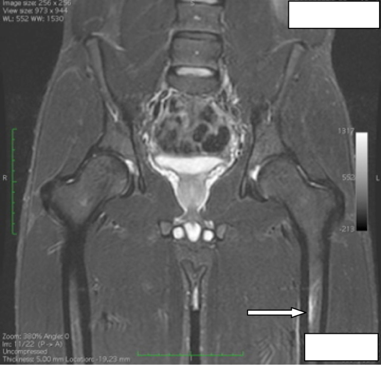
A 19-year-old UK-based Caucasian male soldier reported the onset of progressively worsening bilateral lower limb pain during paratrooper selection training (UK winter months) during a loaded eight mile tactical advance to combat (‘tabbing’) with alternating boot runs. He reported pushing himself to complete the course but was taken off a run due to severe left leg pain. Initial management with entonox and oral non-steroidal anti-inflammatory drugs provided some relief, but symptoms generally worsened over the following few days with an inability to weight bear. Radiographs of the pelvis, left hip and both lower legs were unremarkable. However, pelvic, left hip and bilateral lower limb MRIs demonstrated an evolving stress fracture of the proximal left femoral diaphysis with subcortical bone marrow oedema and periosteal oedema medially and laterally (Figure 1) and a probable chronic stress fracture of the left tibia with bilateral mid-tibial periosteal reaction (Figure 2). Despite 2 months of relative rest he continued to experience left hip pain and had not resumed full weight bearing. He was of mesomorphic habitus and was partially weight bearing on the left leg with the aid of crutches. Both legs were externally rotated, his observed standing lumbar range of motion was full, pain free and unrestricted but prone lumbar segmental stiffness on palpation was present. He walked with a limp and heel weight bearing exacerbated the left femoral pain and Trendelenberg's test revealed poor right side stability. The lower extremities were neurovascularly intact. Passive left hip internal rotation was restricted with left femoral pain at end range; tender points were palpated at the medial and lateral aspects of the patellar borders and medial aspect of the tibias. The bilateral anterior tibial musculature was non-tender. Percussion of the bilateral tibial shafts reproduced exquisite pain, while prone left femoral stretch reproduced femoral shaft pain. He described persistent left hip pain rated as 8.5/10 on the visual analogue scale (VAS) with partial weight bearing, and bilateral tibial pain with a VAS 4.5/10 on palpation but not with weight bearing. There was no family history of metabolic or endocrine problems. Haematological and biochemical investigations identified a borderline low serum vitamin D level of 49 nmol/L (normal range 50–150 nmol/L). Following 1 month supplementation with calcichew D3 Forte two tablets once daily (20 μg, colecalciferol equivalent), the patient stopped analgesic medication, resumed work and returned to pain free full weight bearing (VAS 0/10). His serum vitamin D level increased by 10% to 54 nmol/L.

An evolving stress fracture (arrow) of the proximal left femoral diaphysis with subcortical bone marrow oedema and periosteal oedema medially and laterally.

Probable chronic stress fracture of the left tibia (arrows) with bilateral mid-tibial periosteal reaction.
Discussion
Vitamin D is essential for normal bone homeostasis. This naturally occurring nutrient is present in few foods with the main source of vitamin D via cutaneous synthesis. Solar ultraviolet radiation varies with latitude, and in winter months at latitudes above and below 37° North or South (London is 51° North) sunlight is insufficient to induce cutaneous synthesis of vitamin D.8 Thus, for geographic reasons UK-based military personnel may become seasonally dependent on bodily stores of vitamin D to maintain bone integrity, which diet alone may be inherently insufficient to sustain.
The Scientific Advisory Committee on Nutrition provides evidence of low vitamin D status among all age groups living in the UK.9 Other factors such as the use of sunscreens, lighter skin pigmentation, occupational deprivation of sunlight (eg, submariners), deliberate avoidance of the sun and the wearing of protective military clothing may also interfere with cutaneous vitamin D synthesis.10–12 The UK military population, with its unique, physically demanding roles and special protective clothing and equipment requirements limiting sun exposure, may be at an increased risk of vitamin D deficiency associated lower limb stress fractures.
Though calcichew was prescribed, calciferol preparations (Dekristol) available in the UK may be more appropriate for treating deficiency states.13 According to the Defence Medical Rehabilitation Centre who use the Imperial College of Endocrinology DeKristol Guidelines, a loading dose over 3 months of colecalciferol (Dekristol) 20 000 IU capsules (one capsule weekly for 12 weeks) followed by a maintenance dose of one capsule every 2 weeks is recommended. Higher doses may be required if the patient is taking drugs that accelerate vitamin D metabolism or if there are concerns regarding absorption. Maintenance should be continued so long as risk factors for vitamin D deficiency are present. Vitamin D supplementation appears safe14 and has been shown to facilitate recovery from these injuries.15
Few intervention studies prescribe calcium and vitamin D supplementation to military recruits as a preventative measure. Lappe et al4 reported calcium and vitamin D supplementation substantially reduced the incidence of stress fractures by 20% in female naval recruits. This agreed with a study of Finnish male recruits16 which concluded that vitamin D supplementation may lower the serum parathyroid hormone levels and so possibly reduce the incidence of stress fractures. However, Schwellnus and Jordaan17 reported conflicting results and found no statistically significant effect of large-scale calcium supplementation (500 mg/day) beyond usual dietary intake in preventing stress fractures in male recruits during a 9-week physical training programme.
Conclusions
Sunlight exposure and dietary intake may be inadequate to maintain normal serum vitamin D levels in some UK-based military personnel. Soldiers presenting with stress fractures may have unrecognised hypovitaminosis-D which, if left untreated, may delay fracture healing. This case suggests that prescribing calcichew or calciferol may help with fracture healing. Furthermore, the vitamin D status of military personnel in the UK is unknown, yet there is evidence to suggest that vitamin D status is low among all age groups in the UK; therefore, large scale intervention studies of vitamin D supplementation to possibly reduce the incidence of stress fractures in military personnel might be warranted.
References
Footnotes
-
Acknowledgements Lt Col Rhodri Phillip, Consultant, Rheumatology & Rehabilitation and Mrs Sarah-Jane King, Clinical Specialist Physiotherapist.
-
Contributors JI identified and managed the case. JI and RD diagnosed and treated the patient and they are guarantors. JI, RD, MG and MT drafted and revised the paper.
-
Competing interests None.
-
Patient consent Obtained.
-
Provenance and peer review Not commissioned; externally peer reviewed.