Article Text

Abstract
Introduction Ballistic gelatin is the most common tissue simulant used to reproduce the penetration of projectiles into muscle but published data to support its use are primarily based on bullets, despite explosive fragments being the most common cause of injury to soldiers on current operational deployments. Published ballistic tests using animal and artificial skin and muscle tissue surrogates also lack standardisation in methodology such that limited comparisons with that of human tissues can currently be made.
Method Three masses of cylindrical NATO standardised fragment simulating projectiles (FSPs) were fired at 20% ballistic gelatin and the hind thighs of a killed goat. Threshold (Vth) and V50 velocities required for skin perforation and depth of penetration (DoP) into muscle were compared with gelatin. The intercept and gradient of the linear regression lines for DoP versus velocity were compared between gelatin and goat with significance defined as p<0.05.
Results V50 goat skin perforation velocities for the 0.16, 0.49 and 1.10 g FSPs were 121.1, 103.7 and 97.8 m/s, respectively. There was a significant difference in the V50 required to perforate the gelatin surface compared with goat skin for the 0.16 and 0.49 g FSPs but not the 1.10 g. There was no statistical difference in the gradients for DoP versus velocity between animal and gelatin for either the 0.16 or 1.10 g FSPs.
Discussion This study has produced data for skin perforation velocities and generated algorithms describing velocity versus predicted DoP into muscle for three standardised projectiles, which will be used to improve the fidelity of future injury models. 20% gelatin was demonstrated to accurately reproduce the retardation of the 1.10 g FSPs into goat muscle but the addition of a skin simulant will be required to accurately predict DoP for FSPs less than 1.10 g.
Statistics from Altmetric.com
Key messages
-
Physical models remain an essential tool in predicting the effects of ballistic injury.
-
Ballistic gelatin is the most common tissue simulant used to reproduce the penetration of projectiles into muscle.
-
Published data validating gelatin are primarily based on bullets, despite explosive fragments being the most common cause of injury to soldiers on current operational deployments.
-
Existing data testing fragment simulating projectiles (FSPs) into animal tissue and simulants are of limited value due to poor descriptions of methodology and lack standardisation.
-
The retardation of a NATO standardised cylindrical 1.10 g FSP into goat muscle was shown to be accurately reproduced by 20% gelatin.
-
A skin simulant will be required to accurately predict retardation of cylindrical FSPs less than 1.10 g.
Introduction
Improvised explosive devices (IEDs) remain the most common cause of injury to UK service personnel in the current military environment;1 however, the majority of research into the penetration of human tissues by ballistic projectiles has focused on bullets.2 ,3 Fragments from IEDs are more commonly irregular in shape, increasing their drag coefficient in air in comparison with more regularly shaped missiles such as spheres.2 Therefore, although the initial velocities of such fragments are usually in the region of 1000–1500 m/s, it is believed that most injuries are from fragments with velocities of less than 500 m/s at the time of impact.4
Physical models remain an essential tool in predicting the effects of ballistic injury and enable comparisons of potential mitigative systems to be made. However, the shapes, sizes and consistency of fragments produced in explosions are not always easy to quantify. Analysis of fragments produced by common explosive weaponry in conjunction with those fragments removed from injured service personnel will give some indication of representative fragments. In an attempt to standardise ballistic fragment testing between countries, a North Atlantic Treaty Organization (NATO) Standardising Agreement (STANAG) was published in 2003 describing reproducible fragment simulating projectiles (FSPs) of fixed dimensions, masses and shapes.4 Most previous experiments using FSPs have used spheres5 due to their greater reproducibility despite the recognition that cylinders are generally more representative of IEDs.2 ,6 ,7
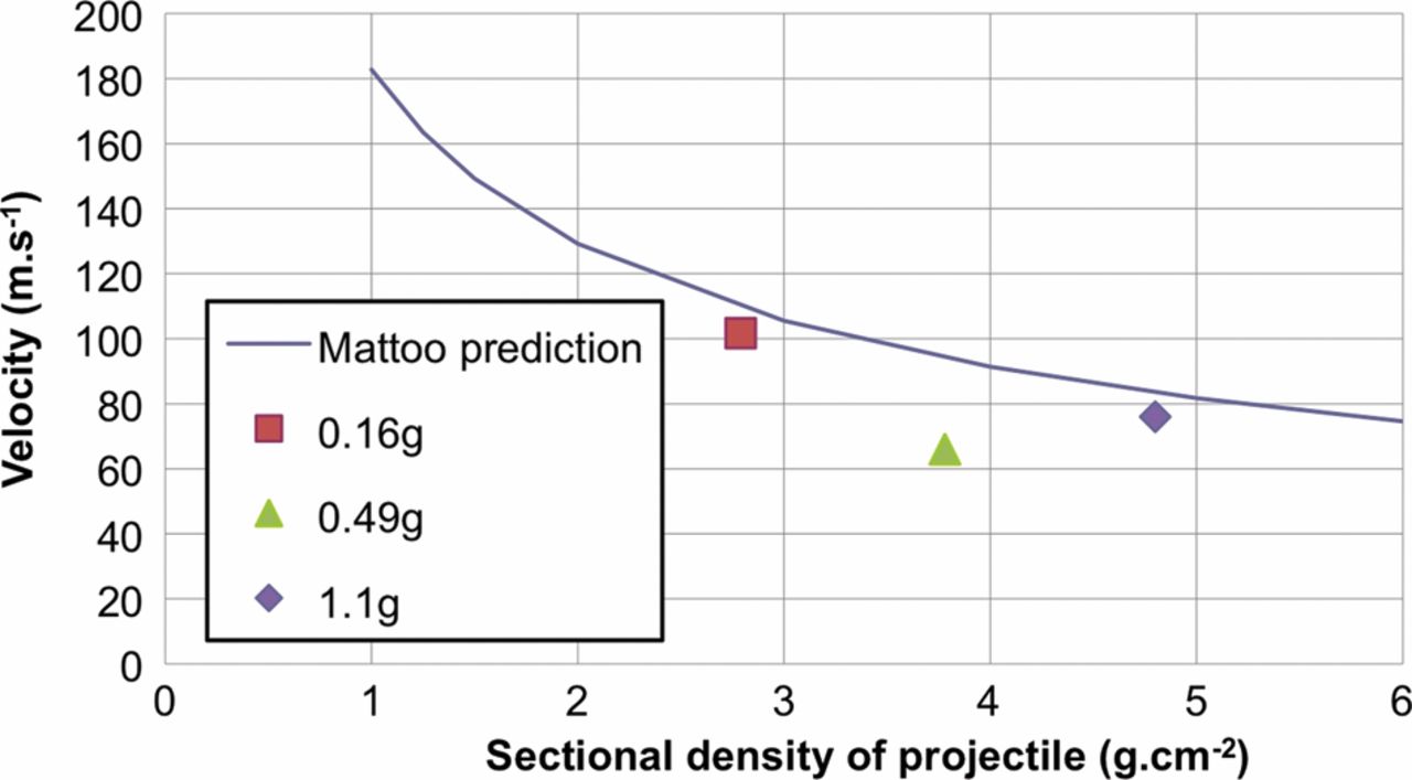
Our knowledge of the resistance of human tissues to explosive fragments is based upon tests on postmortem human subject (PMHS), animal and physical models. PMHS literature has focused more on the ballistic properties of skin rather than muscle. However, the age range of PMHS is typically older than the population of interest when developing Personal Protective Equipment (PPE) (usually healthy young men). Multiple authors5 ,8––10 have published equations based on PMHS studies to predict the velocity required for differing sizes of bullets and air rifle pellets to penetrate and perforate skin. These equations differ from one another primarily because they are based on a very small data set and use different methods to determine skin perforation. Some papers use threshold velocity (Vth) (the minimum velocity in that experiment which resulted in skin perforation) whereas other papers use V50 velocity (the velocity at which 50% of projectiles perforate skin). Other inconsistencies include a lack of description of what is actually meant by skin perforation as well as inadequate descriptions of the dimensions and masses of the projectiles used. For example, in the UK skin ‘perforation’ means that the projectile has passed through all layers of skin, but the term ‘penetration’ is used to describe this in the USA. Sectional density (the mass over cross sectional area) of the projectile is a way of standardising projectile characteristics; it has been shown to be a good predictor for skin perforation and is believed to account for all projectile geometries, sizes and densities.8 ,11 Jussila et al11 felt that Mattoo’s8 suggested empirical relationship between Vth for skin perforation versus projectile sectional density (Figure 1) was the best fit for the limited existing evidence.

Mattoo's suggested empirical relationship8 relating Vth (threshold velocity, m/s) for skin perforation versus projectile sectional density (S=mass over presented area, g/cm2).
Artificial substitutes for skin and muscle have been sought to allow greater reproducibility between testing and to alleviate the need for PMHS and animal testing. Gelatin is generally accepted as a good substitute for modelling animal muscle.5 ,12––14 In turn, it is widely accepted that animal muscle is a good analogy for human muscle, despite no direct comparisons ever having been published in the open literature.2 ,5 Although missile tracks in gelatin blocks are not direct indicators of the amount of tissue damaged by penetrating projectiles, gelatin does simulate the average tissue response in terms of projectile penetration depth and relating temporary cavity volume to energy deposition.4 Despite considerable research,11 no practical artificial skin simulant has been found, generally relating to the difficulties in the consistent reproducible production of the proposed materials.5
Due to the scarcity of appropriate PMHS in conjunction with limitations in the ballistic properties of artificial substitutes, there has been an intermittent need for animal surrogates, particularly in the modelling of human skin. The choice of animal appears to have been limited to that of goats, pigs and dogs.5 Dogs are believed to be a poor substitute in modelling ballistic trauma in humans3 and the few published results in the literature lack conformity due to the great variation in skin properties between breeds.15 Porcine muscle is believed to be a good analogy of human muscle for ballistic testing;5 ,13 ,16 however, the published evidence consists of a single paper,4 which showed good correlation between pig muscle and 20% gelatin at higher velocities but poor correlation at lower ones (<500 m/s). Pig skin is coarser and thicker than human skin,17 reflecting significantly different biomechanical properties, especially in terms of elasticity and tensile strength due to the greater density of collagen fibres;17 it is therefore accepted to be a poor substitute for human skin in terms of ballistic testing.5 ,18
Goat skin has been the most traditional animal surrogate for modelling the penetrating effects of ballistic impacts into human skin10 ,19 ,20 due to perceived similarities in biomechanical properties.5 ,10 ,20 ,21 Goat skin has the same three layers as human skin, namely epidermis, dermis and subcutaneous tissue22 ,23 and also varies in thickness in different body areas, being deepest on the dorsal aspects.24 The thigh is the body area that has traditionally been used for ballistic penetration testing, most likely because it represents a large surface area to increase the number of available shots and has a large proportion of muscle. The thickness of human thigh skin has been described as being between 1.5 and 1.9 mm,25 ,26 thinner than the 3–4 mm described in ballistic experiments on goats.10 ,20 However, the measurements on goat skin used that separated from the underlying muscle and therefore it is unclear if these measurements represent true thickness in vivo.
A systematic literature review including MEDLINE, Google Scholar, Scopus and the Web of Science prior to the start of our experimentation found two pertinent papers in peer-reviewed journals10 ,21 and three open-access reports20 ,27 ,28 (Table 1) pertaining to ballistic penetration of goat skin and/or muscle. In addition, a single historical document was also identified in the newly available declassified documents in the archives of our institution.29 Existing published ballistic experimentation using goats usually pertain to the surgical management of ballistic injury3 and no histopathological data exist regarding its suitability in terms retardation of projectiles. These papers demonstrated a lack of conformity in definitions of penetration, methodology and projectile description such that clear comparisons cannot be made between one another or PMHS data. The aim of this study was to determine the velocity of skin perforation and depth of penetration (DoP) into muscle of three internationally standardised FSPs into intact goat thighs using a clearly described methodology.
Previous published ballistic testing using goats
Method
Three masses of FSP (0.16, 0.49 and 1.10 g) were chosen in accordance with the NATO STANAG.15 The 0.16 and 0.49 g FSPs were standard cylinders and the 1.1 g a chisel-nosed cylinder (Figure 2). The chisel-nosed cylindrical 1.10 g FSP has been the standard projectile for the appraisal of body armour and helmets to date.2 ,10 ,20 ,21 ,30 The 0.16 g FSP was the closest size to that used by Bowyer and colleagues,4 Kokinakis and Sperrazza20 and Sperrazza and Kokinakis10 and is believed to be representative of the most common size of preformed fragmenting munition.10 ,20 ,31 ,32 A third fragment size was chosen half way between the two (0.49 g) on the STANAG sizing and was similar to the 0.54 g chosen by Jussila et al11 and 0.44 g used by Light.21

A 1.10 g chisel-nosed cylindrical fragment simulating projectile: dimensions derived from those specified in the NATO Standardising Agreement.15
Gelatin was prepared using the following standardised method used throughout our institution. Type A ballistic grade (250 bloom, 20% by mass) dry gelatin powder was mixed with distilled water at 70°C±5°C. The water was stirred while the gelatin flakes were added slowly. When all the gelatin was added, it was stirred for an additional 5 min. It was then covered and allowed to stand for 5 min. After this, it was stirred once more for 5 min, and then allowed to stand for a further 45 min. Any excess foam that had formed on the surface of the gelatin was scraped off and the liquid gelatin decanted into moulds. Following cooling to 20°C, the gelatin block was removed from the mould (dimensions 45 cm×20 cm×20 cm) and stored at a temperature of 10°C±2°C for 8–12 h.
The goat used was a 4-year-old Saanen breed (Capra hircus) weighing approximately 60 kg. Ethical approval had been obtained from the Ministry of Defence Research and Ethics Committee. The animal was killed humanely using a Schedule 1 method and had its hind legs clipped to remove any hair in accordance with previous experiments.9 Ballistic testing started within 15 min of the animal being killed. Each leg was elevated in turn using rope until the leg was taut (but not tight) and shots aimed at the thigh to provide comparisons with previous experiments.10
The animal or gelatin blocks were placed in front of a firing rig, with a 5 m distance between the end of the barrel and the target. FSPs were fired from a Pressure Housing weapon system, with a separate smooth bore barrel for each different diameter projectile. The projectiles were propelled using rechargeable 37 mm compressed air cartridges, using pressures of 3–20 MPa. Velocity was measured using solid state velocity equipment with a 1 m separation between the velocity heads. The V50 (the velocity at which 50% of projectiles perforate skin) was determined to represent the most mathematically accurate and reproducible method of describing perforation and allowed comparison with results by Sperrazza and Kokinakis10 and Lewis et al.27 The Dstl Critical Perforation Analysis tool is a graphical user interface based on the statistical software package ‘R’.33 This software uses probit analysis to calculate both a V50 velocity and a 95% CI for that V50 velocity. It also was used during the trial to show when the 95% CI was sufficiently small to give an accurate and reliable result, at which point the testing with that fragment was stopped. Perforation was determined by a military surgeon and was defined as an FSP that traversed through all the three layers of skin (epidermis, dermis, subcutaneous tissue), but did not cause underlying muscle damage. Non-perforation was classed as anything less than full perforation of the skin such as the FSP bouncing off skin or penetrating a partial thickness of skin without breaking the posterior surface of the skin. Although statistically weaker, the Vth (the minimum velocity in which perforation occurred) was also calculated to allow comparison with Mattoo's paper.8
Shots were fired at the lateral thigh surface of all four limbs and filmed using high-speed video to ascertain if tumbling of the FSP occurred prior to impact. Due to the front legs being smaller than the rear, less shots were fired into the former (approximately 7–10 shots in each leg) than the latter (approximately 10–15 shots in each leg). A laser targeting device attached to the rifle barrel enabled accurate shot placement to within approximately 5 mm, aiming for a minimum distance of 20 mm between skin impact locations at velocities unlikely to perforate skin in an attempt to maximise the number of shots but limit damage to adjacent skin. For those shots at higher velocities, a minimum of 40 mm between entry wounds was attempted to prevent overlapping of the wound tracts. All shots were fired at the posterior aspect of the leg and skin depth (surface of skin to surface of muscle) was measured with callipers at four points on each leg (superior, inferior, medial and lateral).
DoP for each FSP perforating into muscle was determined using a metal rod with graduated measurements. There was some concern that this technique might not be accurate for the deeper penetrations where it was difficult to feel the rear surface of the FSP. In these cases, the metal rod was left in situ and a plain radiograph taken to ensure that the rod was touching the FSP; should it be incorrect, the rod length could be adjusted. DoP versus velocity was plotted for each size of FSP into goat tissue using Microsoft Excel for Mac 2011 (Redmond, Washington, USA) and the gradient and intercept of the line of best fit compared with that of gelatin using a Student t test with a significance of <0.05. Mean DoP for shots into the left legs were compared with those into the right.
Results
Skin thickness was between 3.0 and 3.5 mm (mean 3.2). A total of 77 shots were fired using three sizes of FSPs. Values for V50 and Vth are demonstrated in Table 2. The Vth for each FSP was lower than that predicted by the empirical equation suggested by Mattoo8 (Figure 3).
Skin perforation velocities in relation to dimensions and masses of fragment simulating projectiles

Radiographs of metal markers inserted into wound tracks used to increase the confidence in accurately predicting depth of penetration of fragment simulating projectiles (FSPs); note FSPs lodged under contralateral skin surface.
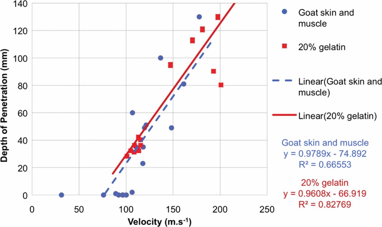
Graphical representation of a linear correlation for the results of DoP versus V50 between goat skin and muscle and 20% ballistic gelatin is demonstrated in Figures 4⇓–6. There was no significant difference in the gradients of the regression lines between the gelatin and animal data for both the 0.16 g (p=0.14) and 1.10 g FSPs (p=0.93). Although there was close correlation between the gradients of the regression lines for the 0.49 g FSP, the gradients were significantly different (p<0.001). There was no significant difference in the intercept of the regression lines between the gelatin and animal data for the 1.10 g FSP (p=0.76). There was a significant difference for the intercept in both cases. No tumbling of FSPs was seen when the high-speed videos of gelatin penetration were reviewed. There was no statistical difference (p<0.05) in DoP measured for shots into the left legs (mean 52 mm) compared with the right (mean 56 mm).

Skin perforation threshold velocity (Vth) versus sectional density (g/cm2) for three standardised fragment simulating projectiles as determined by our experiment, compared with the empirical line of best fit derived from Mattoo's 8 prediction.
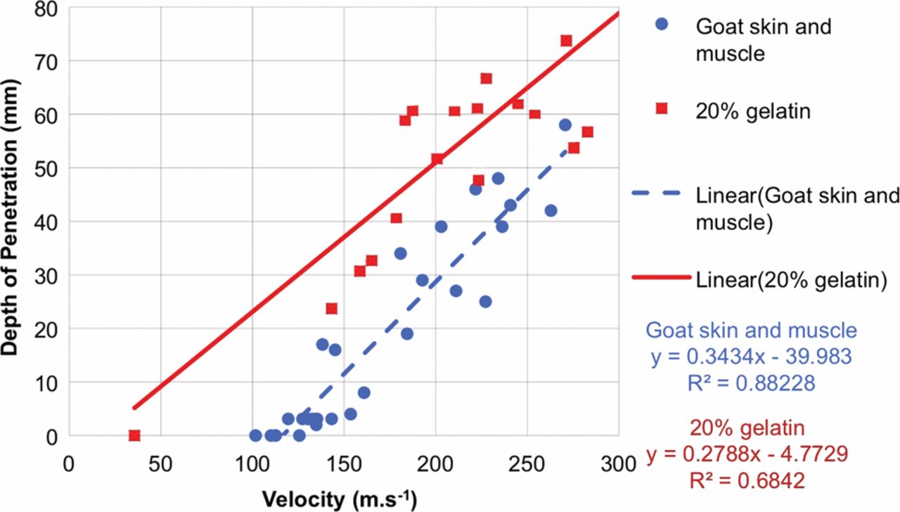
Linear correlation of values relating depth of penetration to V50 velocity for 0.16 g cylindrical fragment simulating projectile fired into both 20% gelatin and goat skin and muscle.

Linear correlation of values relating depth of penetration to V50 velocity for 0.49 g cylindrical fragment simulating projectile fired into both 20% gelatin and goat skin and muscle.
Discussion
The aim of this study was to ascertain values for skin perforation velocity and DoP into muscle using a goat model as a surrogate for humans using standardised variables and a precise methodology. These results could not be ascertained from any of the previous published studies using goats due to a lack of standardisation in methodology. This study has produced clear data for skin perforation velocities and generated algorithms describing velocity versus predicted DoP into muscle for three standardised projectiles which will be used to improve the fidelity of future injury models.
There was a significant difference in the V50 required to perforate skin compared with the surface of the gelatin block for the 0.16 and 0.49 g FSPs, reflecting the importance of skin in retarding these lighter missiles at low velocities. Skin retardation by goat skin was less than found in comparable pig testing,4 confirming their biomechanical differences. There was no significant difference in either the intercept or the gradients for V50 versus DoP between animal and gelatin for the 1.10 g FSP, demonstrating that 20% gelatin is an excellent tissue simulant for goat muscle for this particular FSP. Due to the significant retardation of the 0.16 and 0.49 g FSPs by skin, it is not possible to comment on the gradient of the line of best fit between DoP and V50 to determine if 20% gelatin is a good tissue simulant for goat muscle for these FSPs. Ideally this experiment would be repeated with firings of these two FSPs into goat muscle with the skin removed, as well as 20% gelatin with a validated skin simulant. Such a simulant is also essential for the testing of the skin perforation of low density and non-metallic fragments, the likes of which can be produced by buried explosive devices.34 Existing skin simulants lack standardisation in materials used and availability and we would recommend further research into their development.
The Vth required for skin perforation for all three fragment sizes in our study were less than that predicted by Mattoo's equation8 based on PMHS skin. Although this may represent a significant difference between goat and human skin, we believe that it more likely reflects that Vth is a statistically poor measurement that is subject to high variability. Only a single unrepresentative low velocity shot is required to skew the data unlike the V50 which represents the velocity at which 50% of fragments perforate and which can also be ascribed a confidence limit. V50 measurements were also different from other experiments that used the same measurement and not Vth. The V50 for skin penetration for a 1.10 g fragment was 97.8 m/s, much higher than the 60 and 62.7 m/s found by Sperrazza and Kokinakis10 and Lewis et al27 respectively who used 1.0 g fragments. We believe this reflects that both of their studies used isolated goat skin, which is unlikely to be representative of human skin penetration due to the lack of support provided by the muscle layer beneath it.5 ,11 We would recommend that future studies of skin perforation should also determine the value of V50 instead of Vth, with consideration of calculating the V01 (the velocity at which only 1% of fragments perforated) should a high confidence of non-perforation be required.
Skin thickness in our study ranged from 3.0 to 3.5 mm, similar to the 3–4 mm described in previous ballistic goat skin studies.10 ,20 Skin was shown to be the main component in the resistance of tissues to projectile penetration, and once skin was perforated it travelled very easily through muscle. Many FSPs had enough residual velocity following skin entry to traverse the whole thickness of muscle but not enough to exit through skin. Plain radiographs demonstrated that the fragments lay directly beneath the contralateral skin surface (Figure 7) in an identical manner to that often found at human postmortems.14 The importance of skin in the retardation of lighter FSPs, especially at lower velocities, means that it could be theorised that areas of the body with thicker skin may potentially be more protected against ballistic fragments and future work is required to quantify this effect.
Linear correlation of values relating depth of penetration to V50 velocity for 1.10 g chisel-nosed cylindrical fragment simulating projectile fired into both 20% gelatin and goat skin and muscle.
Significant gaps in our knowledge of the ballistic penetration of skin and muscle to explosive fragmentation still exist. A lack of comparable methodology between papers and limitations in variables highlight the importance of undertaking further research. Although extensive research has been undertaken in the characterisation of the mechanical properties of living human tissues, very little research has been undertaken to characterise that of PMHS tissues.35 Existing PMHS papers have not stated in what manner the tissue was preserved, but it would be highly likely that storage was in formalin, which causes cross-linkages in tissue proteins making tissues stiffer and less elastic.35 ,36 Future research is therefore required to quantify the differences in the mechanical properties between living human skin and PMHS skin to predict how they may behave differently when ballistic fragments are fired into them. If significant differences are found then it would suggest that existing PMHS data may be flawed and that repeating these experiments with newer methods of preserving PMHS skin such as fresh-frozen storage35 is indicated. It has been suggested that hot and wet skin, as experienced by service personnel currently deployed to Afghanistan, is more susceptible to penetration.37 These results have never been quantified by ballistic testing and future testing is required to determine how skin temperature and moisture content affect the perforation velocity for skin of explosive fragments. Finally, the steel cylinder is generally considered the most representative shape of randomly produced explosive fragments.2 ,6 ,7 This is based upon collection of fragments produced by explosive devices in Arena trials and those found embedded in PPE. Further research is required to produce a broader sample of fragments to either justify the use of the cylinder or determine a more representative shape. This will require accurate characterisation of fragments removed from injured servicemen as well as those visualised with radiology.
References
Footnotes
-
Contributors Design: JB and GRJ; Literature review: JB and GRJ; and Manuscript preparation: JB, GRJ and AEH.
-
Competing interests None.
-
Provenance and peer review Not commissioned; externally peer reviewed.